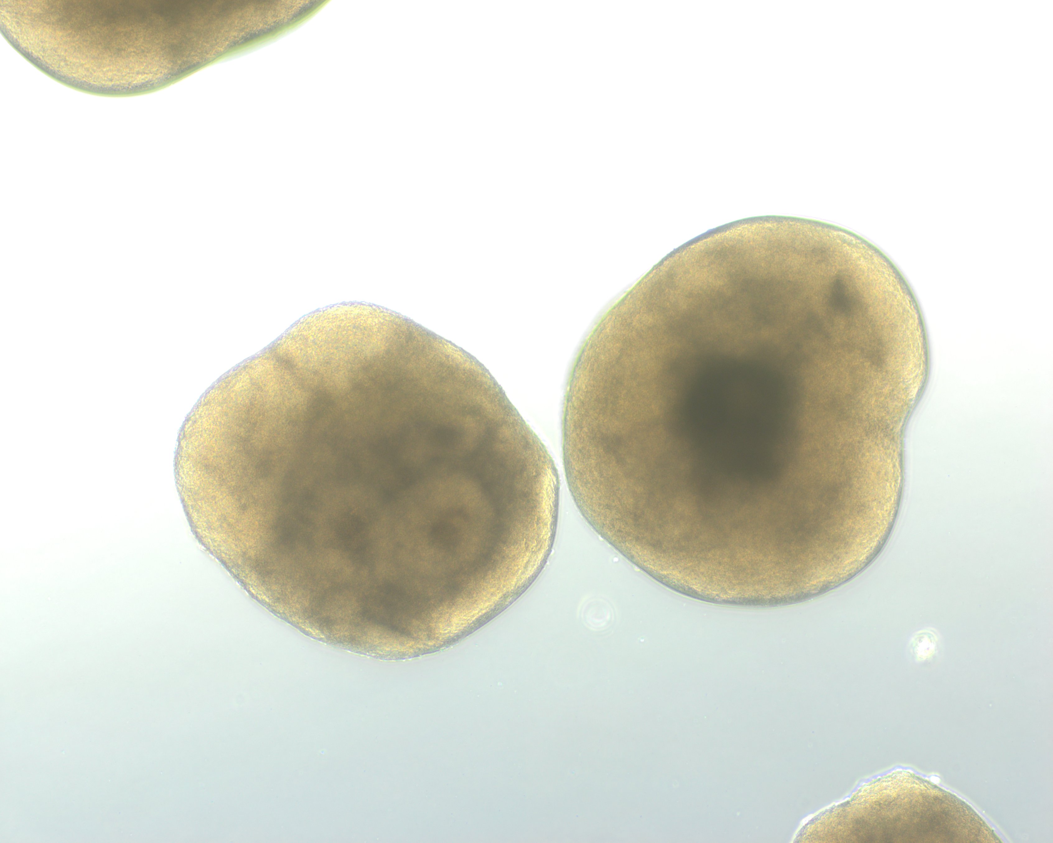
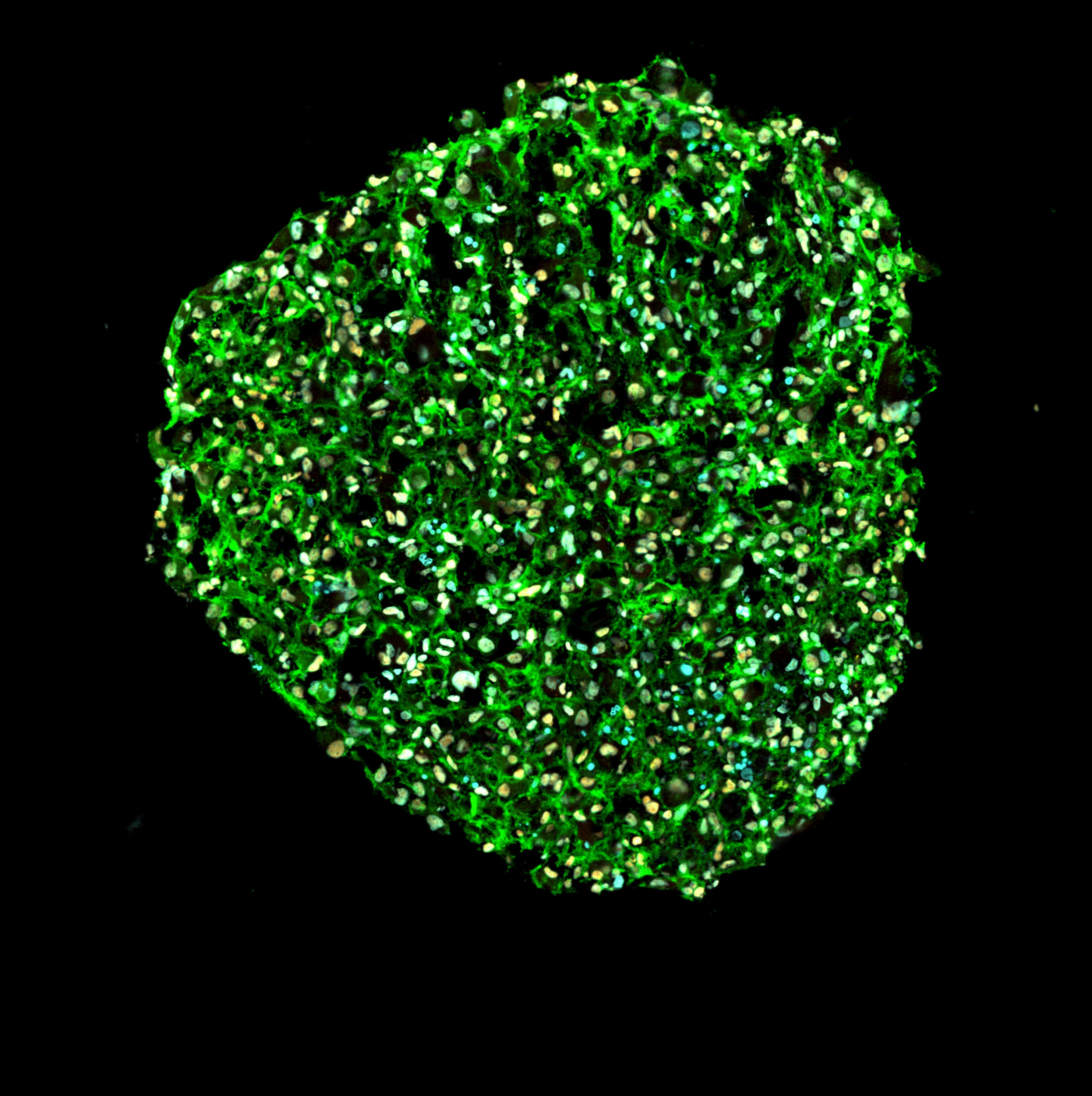
Organoid Models

We develop and apply physiologically relevant human organoid systems for disease modeling, therapeutic testing, and mechanistic studies.

Organoids are advanced three-dimensional (3D) models that mimic the structure and function of human organs. Derived from stem cells, organoids self-organize into complex tissues that closely resemble key features of their in vivo counterparts. By recapitulating aspects of human organ development, physiology, and disease, organoids provide insights that are often difficult to obtain using traditional two-dimensional (2D) cell cultures or animal models. As highly relevant preclinical models, organoids have broad applications in disease modelling, drug discovery and screening, biomarker identification, and predicting patient-specific responses to therapy.

Why Nocteva Organoids

Our Organoid Platform

Key Advantages of Our Organoid Platform

Human-relevant biology
Organoids derived from human iPSCs capture key aspects of human tissue architecture, physiology, and disease.
High reproducibility
Standardized protocols and rigorous quality control ensure consistent organoid generation across batches and studies.
Disease-specific models
Patient-derived organoids enable the study of disease mechanisms and therapeutic responses in a clinically relevant context.
Reliable preclinical platform
More predictive, human-specific data than conventional 2D cultures, complementing or reducing reliance on animal models.
Scalable & assay-ready
Uniform organoids suitable for drug screening, target validation, biomarker discovery, and efficacy testing.
Translational value
Data that support preclinical decision-making and accelerate the path toward clinical development.
Reproducibility & Maturity

Reproducible and rapidly maturing, batch after batch

Our protocols deliver markedly lower batch-to-batch variation than competitors, while reaching human-relevant developmental maturity far faster, for predictable, dependable data.

A
Batch reproducibility (MDS)
Each dot is one organoid batch. Nocteva's batches cluster tightly (highly reproducible), while the competitor's scatter widely.
MDS2
MDS1
Competitor: wide spread
Nocteva: tight cluster
B
Predicted developmental retinal age
Photoreceptor (rod) maturity, inferred from a developmental gene-expression signature.
Maturity →
Adult retina
Competition
Day 210
Nocteva
Day 80
Nocteva
Day 240
Adult
retina
At just day 80, Nocteva organoids already surpass a competitor's day 210 organoids, and by day 240 approach adult-retina maturity.

Shown here for our iPSC-derived retinal organoids as a representative example of the platform's reproducibility.

Platform validation · Retinal organoids as example

How closely our organoids mirror the human organ

Our retinal organoid model illustrates the level of biological complexity and characterization achieved across our platform. It contains all major retinal cell types organized in their native layered architecture, confirmed by brightfield and immunofluorescence imaging. Every model system below is characterized with the same level of depth and rigor.

Brightfield: live morphology
Uniform size and shape, with a visible photoreceptor brush border
Brightfield, uniform organoid batch
Batch uniformity
Consistent size and morphology across the batch
Brightfield, uniform organoid array
Defined structure
Clear internal organization and emerging layers already visible at this stage
Brightfield, single organoid with brush border
Photoreceptor brush border
Dense outer-segment "brush line" at the organoid surface
Immunofluorescence: cell-type composition
Structure and organization of retinal cell types
Immunofluorescence, whole organoid
Whole organoid
CRX⁺ photoreceptors form the outer layer around the organoid
DAPI: nuclei CRX: photoreceptors Calretinin: amacrine / horizontal
Immunofluorescence, layered neural retina
Layered retina
Day 80 retinal organoid showing early stratified retinal organization
CRX: photoreceptor / bipolar ARR3: cones Rhodopsin: rods
3D Imaris quantification of cone and rod photoreceptors
3D Imaris
Rod & cone photoreceptors
3D reconstruction and quantification of mature photoreceptor subtypes
Rhodopsin: mature rods ARR3: cones
Brain Organoids

Brain organoids are iPSC-derived models that recapitulate key features of human brain development and function. Multiple neural cell types self-organize into tissue-like structures, making it possible to study cellular interactions and region-specific biology in a controlled, human-relevant in vitro system.

Nocteva generates both whole-brain (cerebral) and region-specific organoids, a precise platform to investigate disease mechanisms, characterize cellular dysfunction, and evaluate therapeutic interventions where they matter most.

Available region-specific models
Dorsal forebrainVentral forebrainMidbrainHindbrainHypothalamusCerebellumHippocampus
From early morphology to region-specific identity
Midbrain
Ventral midbrain
Midbrain organoid, Day 20 brightfieldDay 20 · BF
Midbrain organoid, Day 50 immunofluorescenceDay 50 · IF
FOXA2THDAPI
Cortex
Forebrain cortical
Cortex organoid, Day 20 brightfieldDay 20 · BF
Cortex organoid, Day 50 immunofluorescenceDay 50 · IF
CTIP2SOX2RELN
Hippocampus
Dorsal hippocampal
Hippocampus organoid, Day 20 brightfieldDay 20 · BF
Hippocampus organoid, Day 75 immunofluorescenceDay 75 · IF
PROX1SOX2
Retinal Organoids

Retinal organoids recapitulate the key features of the human retina, including the presence and organization of all major retinal cell types. Derived from iPSCs, they provide a human-specific platform to study retinal development, disease mechanisms, and therapeutic interventions where animal models and 2D cultures fall short.

Nocteva has optimized retinal organoid generation for robust reproducibility, consistent cellular composition, and low batch-to-batch variability, with standardized differentiation protocols and rigorous quality control.
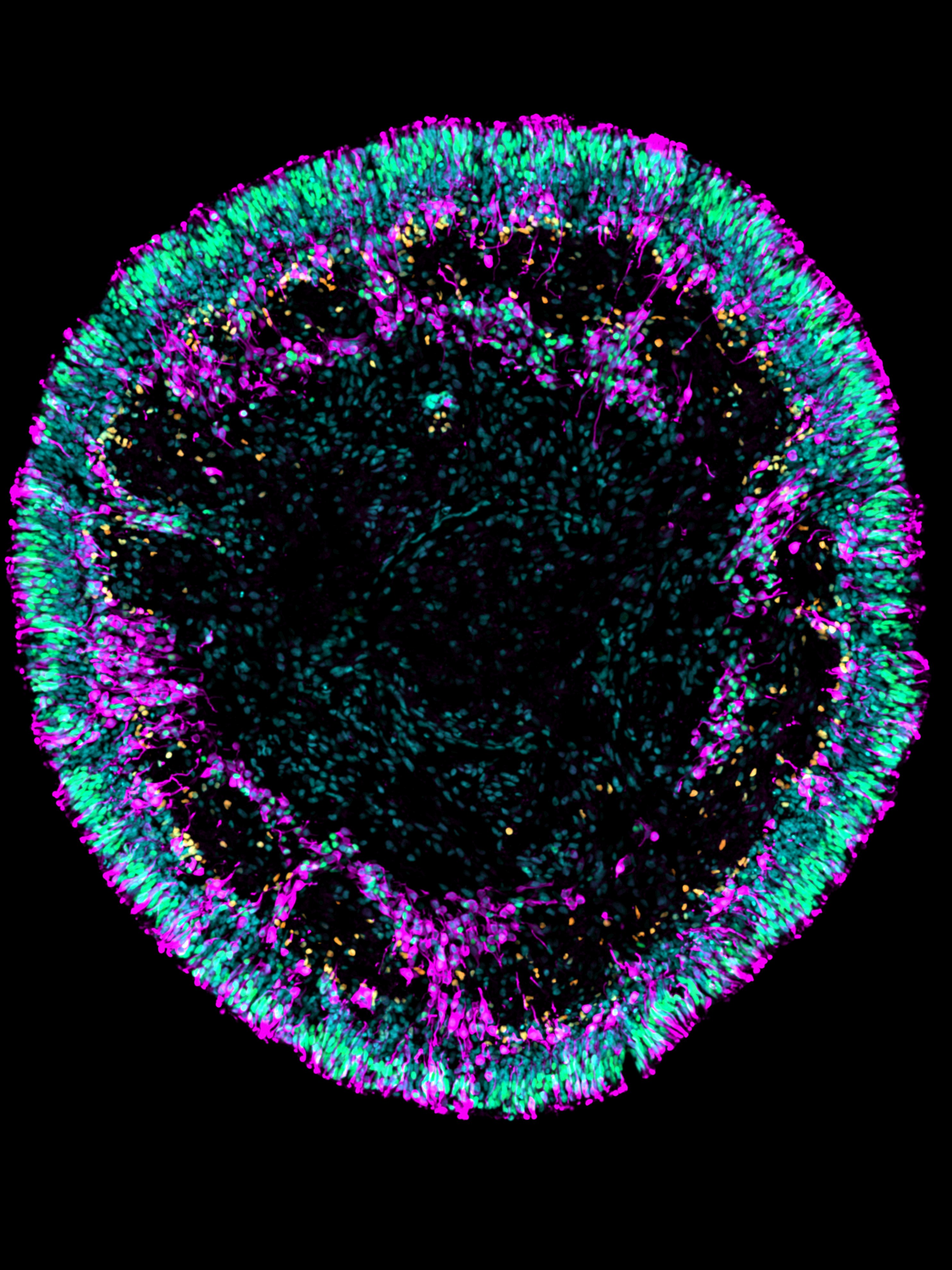
Retinal organoid immunofluorescence, cones, rods, amacrine cells
Photoreceptors & interneurons
Distinct cone and rod photoreceptor populations alongside amacrine interneurons
ARR3 — cone photoreceptors NRL — rod photoreceptors AP2α — amacrine cells Nuclei
Retinal organoid immunofluorescence, rods, bipolar, ganglion cells
Layered retinal circuitry
Rod photoreceptors, bipolar interneurons, and ganglion cells in native-like organization
RHO — rod photoreceptors CHX10 — bipolar cells & progenitors RBPMS — ganglion cells Nuclei
High-magnification detail
Retinal organoid, high-magnification detail
Resolved cellular organization
At high magnification, distinct photoreceptor populations and interneuron processes are clearly resolved within an ordered outer layer — direct evidence of the structural fidelity and cell-type diversity Nocteva's retinal organoids achieve.
RHO — rod photoreceptors CHX10 — bipolar cells & progenitors RBPMS — ganglion cells Nuclei
Applications
Inherited retinal disease modeling
Model retinitis pigmentosa and other degenerations in patient-derived organoids.
Drug efficacy & toxicity screening
Test candidate compounds for efficacy and retinal safety in a human system.
Gene & cell therapy development
Evaluate AAV delivery, gene correction, and cell-replacement strategies.
Biomarker & mechanism discovery
Uncover disease mechanisms and translational biomarkers of response.
Cardiac Models

Nocteva's cardiac platforms span 2D cardiomyocyte monolayers to 3D cardiac organoids — human iPSC-derived models that recapitulate cardiac tissue composition, contractile activity, and electrophysiology for disease modeling and therapeutic evaluation.

Cardiac organoid generation is optimized and standardized for high reproducibility, consistent tissue composition, and robust functional performance.
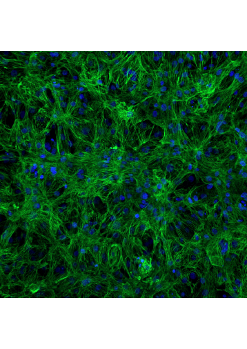
2D cardiomyocyte monolayer
Cardiac monolayer, immunofluorescence
TNNT2 — cardiomyocytesDAPI — nuclei
Live imaging — spontaneous beating
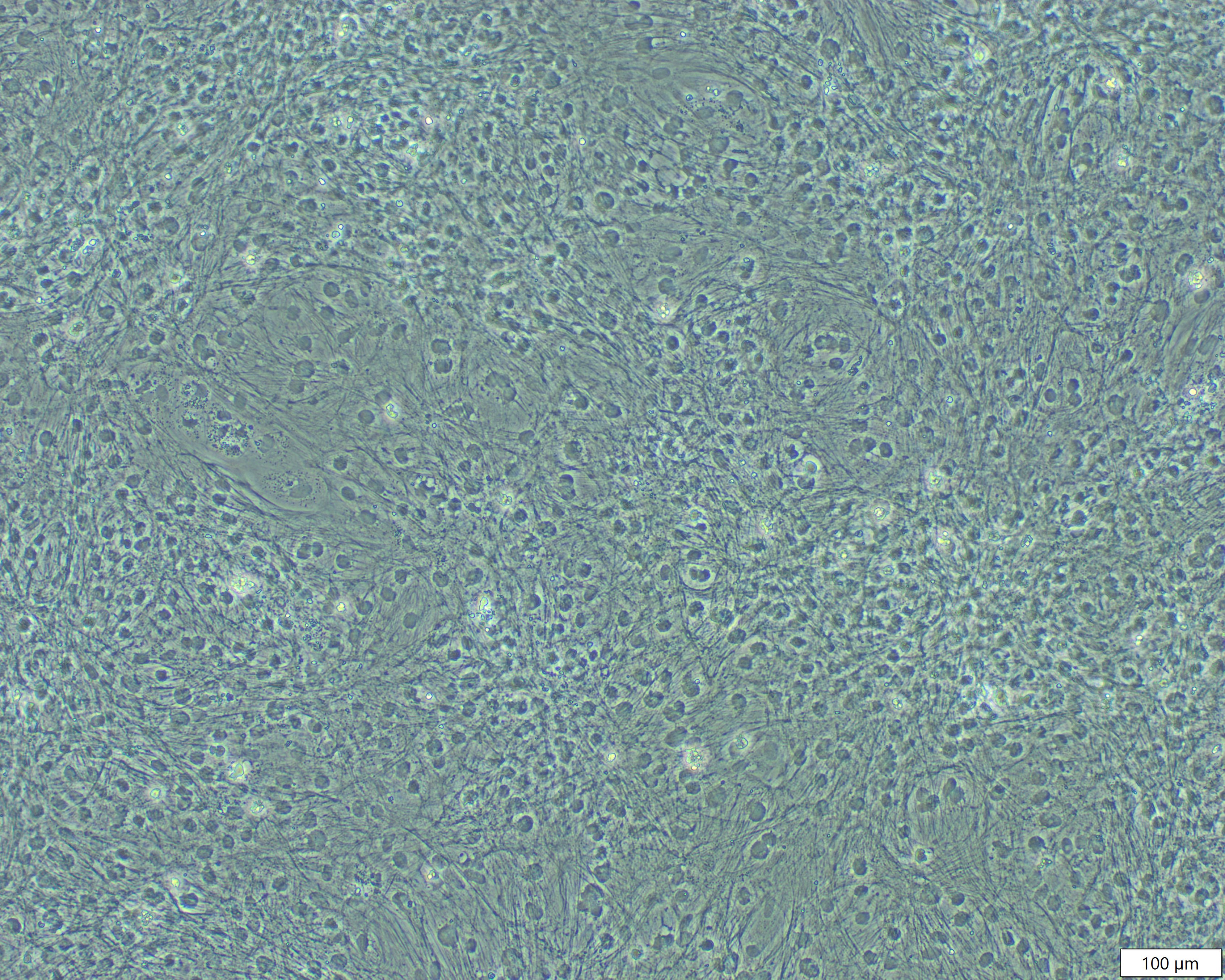
Cardiac monolayer, brightfield
Brightfield — uniform monolayer morphology

iPSC-derived cardiomyocytes form spontaneously contracting 2D monolayers — a simple, scalable platform for cardiotoxicity screening and functional assays.

Cardiac organoids
Cardiac organoid, immunofluorescence
TNNT2 — cardiomyocytesCD31 — endothelial cellsDAPI — nuclei
Calcium imaging — live signaling during contraction
Cardiac organoid, brightfield
Brightfield — uniform, well-defined organoids
Live functional imaging

Live imaging captures rhythmic, spontaneous beating in real time — a direct functional readout of cardiac tissue health, drug response, and electrophysiological activity.

Beating monolayer
Spontaneous, synchronized contraction across a 2D cardiomyocyte sheet.
Monolayer — close-up view
A closer field of view resolving individual beating cardiomyocytes.
Beating organoid
Coordinated, tissue-level contractile activity within a 3D cardiac organoid.
Organoid — alternate view
Consistent, reproducible beating across the organoid.
Neuromuscular Models

Nocteva builds a connected suite of human iPSC-derived neuromuscular models, from neuromuscular, skeletal muscle, and spinal cord organoids to functional spinal motor neurons, providing physiologically relevant platforms to study motor-neuron and muscle biology, disease, and therapeutic response.
Spinal cord organoids
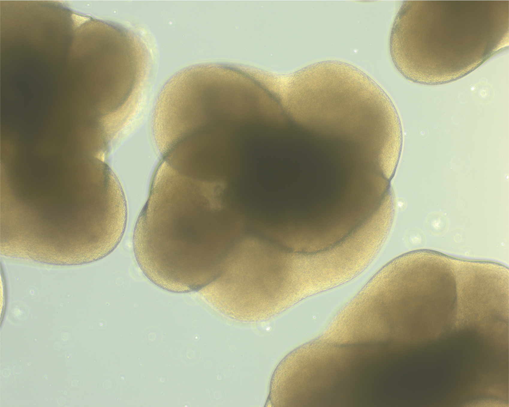
Spinal cord organoid, brightfield
Spinal cord organoid, immunofluorescence

Spinal cord organoids reproduce the cellular diversity and organization of the developing human spinal cord, including populations of motor neurons — a platform for studying motor circuits, spinal development, and motor-neuron disease.

ISL1/2 — motor neurons PHOX2B — motor neurons DAPI — nuclei
Skeletal muscle organoids
Skeletal muscle organoid, brightfield
Skeletal muscle organoid, immunofluorescence
Contractile human muscle tissue
Multinucleated muscle fibers, tissue organization, and contractile function.
TITIN — sarcomeric marker
Neuromuscular organoids
Neuromuscular organoid, brightfield
Neuromuscular organoid, immunofluorescence
Functional neuromuscular junctions
Motor neurons, muscle cells, and terminal Schwann cells self-organize into functional neuromuscular junctions.
TUJ1 — motor neurons Fast myHC — muscle cells GFAP / S100b — Schwann cells αBTX — AChR clusters
Spinal motor neurons · 2D culture

iPSC-derived spinal motor neurons form dense, interconnected networks and mature into cholinergic neurons — enabling the study of motor-neuron biology and diseases such as ALS and spinal muscular atrophy.
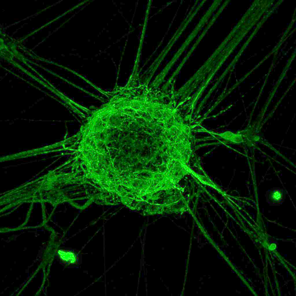
Spinal motor neurons, MAP2 immunofluorescence
Mature neuronal networks
Neurons interconnect and form spherical structures with extensive dendritic arbors
MAP2 — mature neurons & dendrites
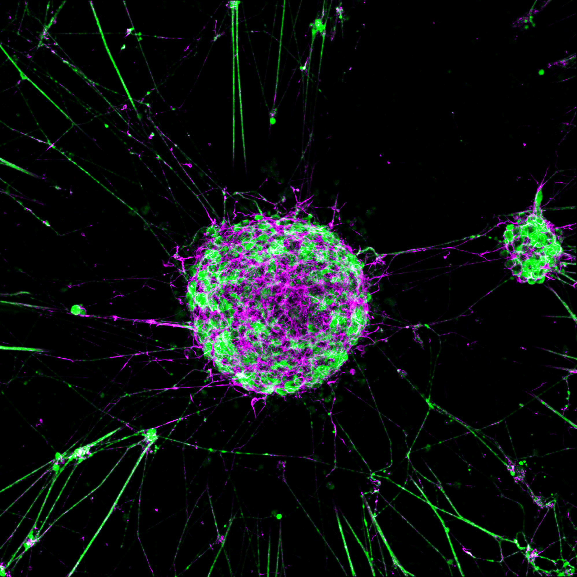
Spinal motor neurons, ChAT, actin, DAPI immunofluorescence
Cholinergic identity
ChAT⁺ motor neurons — the enzyme that synthesizes acetylcholine for muscle activation
ChAT — cholinergic motor neurons Actin — cytoskeleton DAPI — nuclei
Lung Organoids

Lung organoids are iPSC-derived models that recapitulate key features of human airway and alveolar tissue, including epithelial organization and cell-type diversity, providing a human-relevant platform for studying lung development, disease, and therapeutic response.

Nocteva's apical-out lung organoids present the epithelial surface outward, mimicking the in vivo interface bacteria encounter — making them particularly suited for modeling bacterial infection and testing antibiotics.
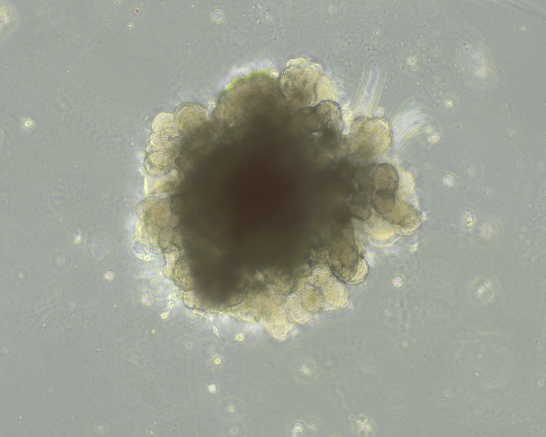
Lung organoids, brightfieldBF
Live morphology — lung organoids in culture
Lung organoids, immunofluorescence overviewIF
Overview — epithelial organization across organoids
Apical-out lung organoid, immunofluorescence close-upIF
Apical-out organoid — ciliated surface facing outward
Acetylated α-tubulin — cilia Actin — cytoskeleton DAPI — nuclei
Applications
Bacterial infection modeling
Apical-out epithelium exposed to pathogens under in vivo-like conditions.
Antibiotic & therapy testing
Evaluate antimicrobial efficacy on infected human airway tissue.
Patient-Derived Tumor Organoids

Patient-derived tumor organoids, including high-grade glioma and glioblastoma (GBM), are generated from patient tumor material and preserve the cellular heterogeneity, invasive behavior, and molecular signatures of the original tumor.

Nocteva establishes and characterizes tumor models for mechanistic studies, therapy testing, and biomarker discovery, with matched molecular profiling available through our multi-omics services.
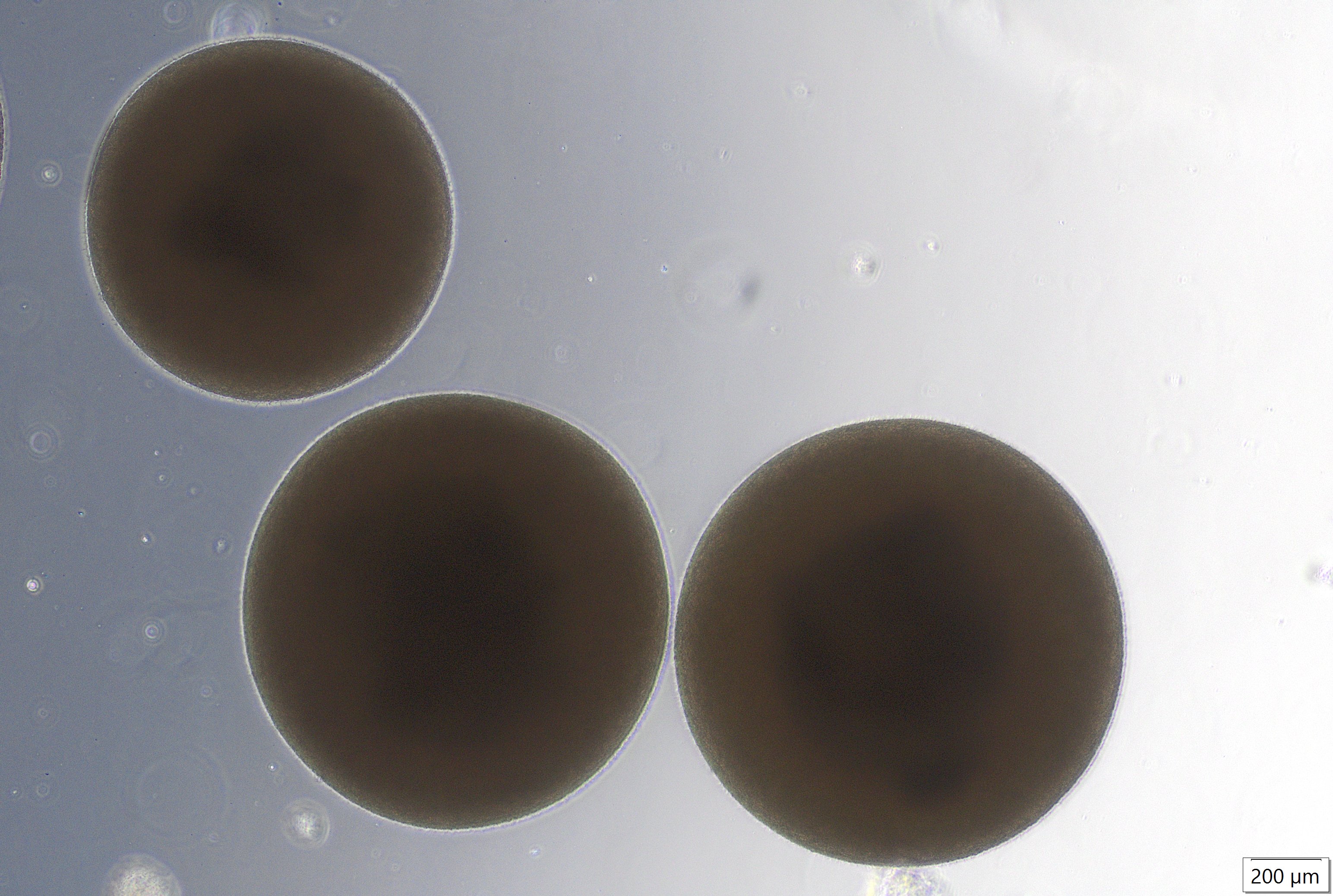
Glioblastoma organoid, brightfield, day 26Day 26 · BF
Live morphology — glioblastoma organoid (GBO) in culture
Glioblastoma organoid, immunofluorescence, EGFR and SOX2IF
Tumor cell populations — EGFR⁺ tumor cells and SOX2⁺ progenitor-like cells
EGFR — tumor cells SOX2 — progenitor-like cells
Applications
Tumor biology & invasion
Proliferation, heterogeneity, and invasive behavior in a patient-relevant 3D system.
Therapy response testing
Small molecules, biologics, and combination regimens on patient-derived models.
Multi-omics characterization
snRNA-seq, proteomics, and epigenomic profiling of tumor models and treatment response.
Biomarker discovery
Molecular signatures of sensitivity and resistance to guide therapeutic development.

Contact

Interested in our services?
We collaborate with researchers, clinicians, and patient communities to drive innovation where it’s needed most.
Your submission has been received!
Oops! Something went wrong while submitting the form.